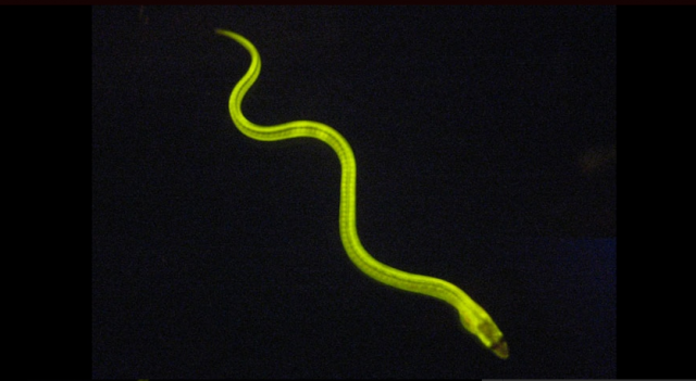
Researchers have discovered a fluorescent protein in a Japanese eel consumed as a popular sushi snack. Amongst other applications, the discovery could help provide a simpler and more sensitive test to detect jaundice and other diseases.
The idiom “seeing is believing” is what drives many biologists to use fluorescence microscopy, where specifically tagged proteins glow when a laser is shone on them. This glow allows researchers to observe phenomena inside cells at very minute scales (at some billionths of a meter).
The importance of one class of proteins that are used as tags, called green fluorescent proteins (GFPs), was recognised by the 2008 Nobel Prize in Chemistry. But so far, all the GFPs have been derived from non-vertebrate animals—those that lack a spinal cord—such as jellyfish and corals.
The discovery of the new fluorescent protein, named UnaG after the Japanese eel unagi, is important not just because it comes from a vertebrate, but also because it differs from any of the fluorescent proteins currently available.
A special glow
The first GFP was discovered in a jellyfish called Aequorea victoria almost 50 years ago. Since then, tiny tweaks to this GFP and a few others that were discovered later on have given researchers a set of reliable tools to help them probe how a cell works (many of the tweaks have gotten the protein to glow in a rainbow of colors). By attaching GFP to the proteins they’re interested in, scientists can pinpoint and study individual proteins among thousands that are at work in a cell.
The discovery of a new fluorescent protein was recently published in the journal Cell. A team led by Atsushi Miyawaki at the RIKEN Institute in Japan found UnaG when they were studying the muscle fibers of freshwater eels. The protein is unique not just because it is the first found in a vertebrate, but also because of the way it fluoresces. In most GFPs, the chromophore (the part of the molecule that can absorb and emit light) is part of the protein itself. UnaG glows by integrating a molecule from outside the protein.
This molecule turns out to be bilirubin, which is present in eel muscles but is also formed when haemoglobin breaks down in human blood. Levels of bilirubin have been used for decades to assess liver health and diagnose diseases such as jaundice. So this feature of UnaG gives it the potential to detect bilirubin and act as an indicator for liver malfunction.
Binding to bilirubin gives UnaG some additional properties that no other GFP currently has. First, since its chromophore isn’t part of the protein itself, it is only half the size of current GFPs, which makes it easier to use it to tag proteins without interfering with their function. Second, most GFPs must react with oxygen to produce their chromophore and thus become fluorescent. UnaG does not. This means it could enable the illumination of cells in tissues where oxygen is scarce, such as some cancerous tumours.
Biology has always been driven by the ability to “see” nature. Modern tools have allowed scientists to see beyond what the naked eye could offer. And the race is on to see phenomena happening on ever smaller scales. UnaG is a promising step in that direction.![]()
Cell, June 2013. DOI: 10.1016/j.cell.2013.05.038 (About DOIs)
Luc Henry is postdoctoral fellow at the Swiss Federal Institute of Technology in Lausanne.
This article was first published at The Conversation.
reader comments
12